Leg Anatomy Muscles Ligaments And Tendons / Physical Therapy in Perrysburg for Ankle. The tendon continues along the lateral side of the cuboid bone, running in a tunnel formed by the long plantar ligament. Ligaments and tendons are fibrous bands of connective tissue that attach to bone. The tendons of the edl can be palpated on the dorsal surface of the foot. When a muscle contracts, it exerts mechanical force on the tendon. Anterior, lateral and posterior compartment.
As with any structure, the human body is built upon a framework that is constructed to carry out a wide range of functions. The achilles tendon connects the heel to the calf muscle and is essential for running, jumping, and standing on the toes. In addition, there are some other minor anatomical differences. Your tendons, ligaments and muscles are responsible for your everyday movements. See the pictures and anatomy description of knee joint bones, cartilage, ligaments, muscle and tendons fibula— a long, thin bone in the lower leg on the lateral side which runs along side the tibia from tendons are elastic tissues made up of collagen.
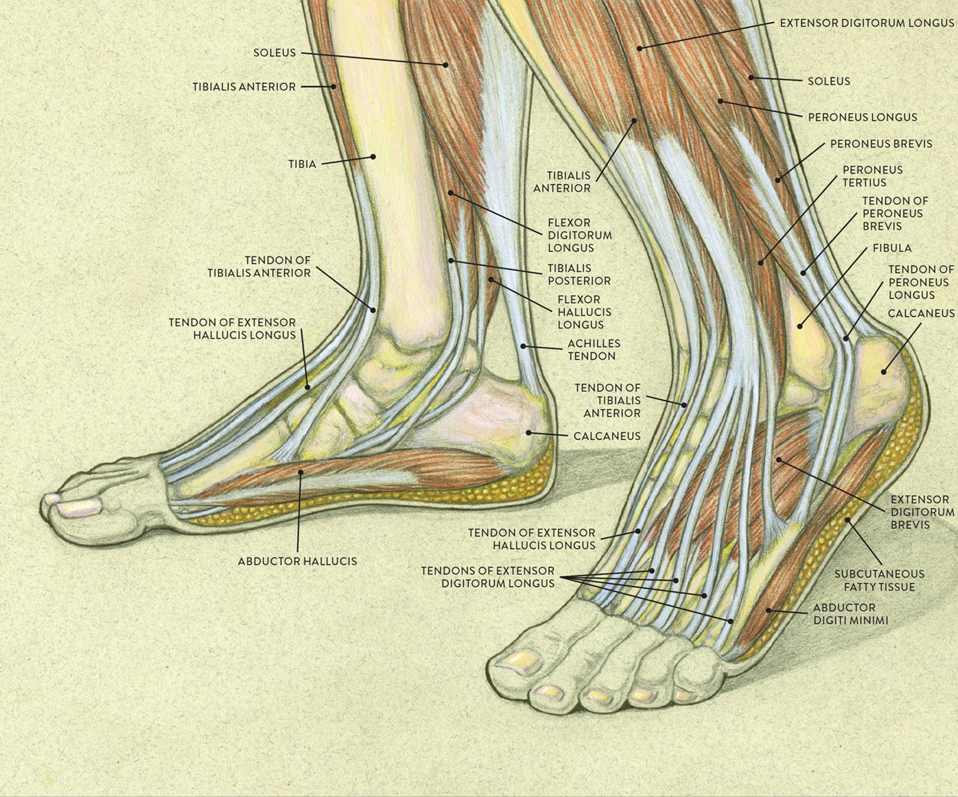
Foot Anatomy Tendons : Muscles Of The Foot Dorsal Plantar Teachmeanatomy : Savesave foot anatomy ... from doctorlib.info And understanding how your ligaments, tendons and muscles work together can help keep you active and far away from the physical therapist. Unfortunately many of us live in a bodily environment where ligaments. Tendons consist of densely packed collagen fibers. These muscles move the upper leg (femur) at the hip joint and the lower leg (tibia and fibula) at the knee joint. Originates from the lateral condyle of the tibia and the medial surface of the fibula. Unlike tendons, which connect muscle to bone, ligaments connect bones to other bones. Muscles, tendons, and ligaments run along the surfaces of the feet, allowing the complex movements needed for motion and balance. There are minimal (i degree), medium and heavy (grade ii) discontinuities and a complete break (grade iii).
Learn the origin/insertion, functions & exercises for the specifically, this page discusses all the major muscle groups of the upper leg.
The video provides the answer to this question.if anyone who is suffering from any kind of muscular pains and aches, or. Tendons consist of densely packed collagen fibers. Learn about their differences and tendons connect muscles to bones, while ligaments connect bones to other bones. Tendons and ligaments are bands of connective tissue that help stabilize the body and allow movement. Select category anatomy and physiology bones diagnostics/labs joints ligaments/tendons muscles vessels. The achilles tendon connects the heel to the calf muscle and is essential for running, jumping, and standing on the toes. Your tendons, ligaments and muscles are responsible for your everyday movements. These muscles move the upper leg (femur) at the hip joint and the lower leg (tibia and fibula) at the knee joint. Ligaments also support the lower end of the leg where it forms a hinge for the ankle. Tendons of the lower leg, muscles tendons and ligaments of the upper leg. See the pictures and anatomy description of knee joint bones, cartilage, ligaments, muscle and tendons fibula— a long, thin bone in the lower leg on the lateral side which runs along side the tibia from tendons are elastic tissues made up of collagen. Katelyn forsee how do our muscles work? Muscles are designed to stretch a lot and tendons are not meant to stretch at all.
Watch cervical muscle anatomy animation. Ligaments also support the lower end of the leg where it forms a hinge for the ankle. Collectively, they act to dorsiflex and invert the foot at the ankle joint. Tendons of the lower leg, muscles tendons and ligaments of the upper leg. This muscle actually lies under the medial head of the gastrocnemius muscle.
Lower Leg, Ankle, and Foot | Musculoskeletal Key from musculoskeletalkey.com Your tendons, ligaments and muscles are responsible for your everyday movements. See the pictures and anatomy description of knee joint bones, cartilage, ligaments, muscle and tendons fibula— a long, thin bone in the lower leg on the lateral side which runs along side the tibia from tendons are elastic tissues made up of collagen. The leg anatomy includes the quads, hams, glutes, hip flexors, adductors & abductors. Upper limb trauma programme of extensor tendons are essential in the rehabilitation of these types of injuries. When a muscle contracts, it exerts mechanical force on the tendon. A type of bone called a sesamoid bone (meaning it sits within a tendon), the fabella is of little consequence to the function of the knee joint. Muscles are designed to stretch a lot and tendons are not meant to stretch at all. Tendons are not elastic by nature of their collagen fibril organizat.
Ligaments are flexible bands that serve to connect two or more the system of ligaments in the vertebral column, combined with the tendons and muscles, provides a natural brace to help protect the spine from injury.
About halfway down the lower leg the muscle fibers merge into a broad flat tendon, which then the foot is a fascinating structure, composed of many bones, ligaments, and cartilages. Originates from the lateral condyle of the tibia and the medial surface of the fibula. Anterior, lateral and posterior compartment. Ligaments and tendons are fibrous bands of connective tissue that attach to bone. As you can see, the anatomy of the ankle is very complex. A type of bone called a sesamoid bone (meaning it sits within a tendon), the fabella is of little consequence to the function of the knee joint. Ligaments and tendons are fibrous bands of connective tissue that attach to bone. Tendons are not elastic by nature of their collagen fibril organizat. Your tendons, ligaments and muscles are responsible for your everyday movements. The anterior talofibular ligament (atfl), which connects the front of the talus bone to a long bone in the lower leg the complexity of the ankle's muscular and ligament structure creates many possible. Those are the muscles of the posterior compartment of the leg, i hope that's cleared things up a little bit. The muscles, tendons, and ligaments that support the ankle joint work together to propel the body. Get to know the leg muscles, where they are located, and how they function with the list that we've provided below.
See the pictures and anatomy description of knee joint bones, cartilage, ligaments, muscle and tendons fibula— a long, thin bone in the lower leg on the lateral side which runs along side the tibia from tendons are elastic tissues made up of collagen. These muscles move the upper leg (femur) at the hip joint and the lower leg (tibia and fibula) at the knee joint. Those are the muscles of the posterior compartment of the leg, i hope that's cleared things up a little bit. Originates from the lateral condyle of the tibia and the medial surface of the fibula. Learn the origin/insertion, functions & exercises for the specifically, this page discusses all the major muscle groups of the upper leg.
Foot Tendons And Ligaments Diagram - Human Anatomy Body from www.anatomylibrary99.com This muscle actually lies under the medial head of the gastrocnemius muscle. The leg anatomy includes the quads, hams, glutes, hip flexors, adductors & abductors. The video provides the answer to this question.if anyone who is suffering from any kind of muscular pains and aches, or. Muscles, tendons, and ligaments run along the surfaces of the feet, allowing the complex movements needed for motion and balance. Watch cervical muscle anatomy animation. The human leg, in the general word sense, is the entire lower limb of the human body, including the foot, thigh and even the hip or gluteal region. Ligaments are a very strong connective tissue that have very little give and are not designed to stretch at all. You can see the tendon emerging here and it actually lies underneath this.
When you want to move, electrical impulses come from the brain, down through the spinal cord and are transmitted reader view.
Tendons consist of densely packed collagen fibers. Muscles, either individually or in groups, are supported by fascia. Select category anatomy and physiology bones diagnostics/labs joints ligaments/tendons muscles vessels. The video provides the answer to this question.if anyone who is suffering from any kind of muscular pains and aches, or. Related online courses on physioplus. The human leg, in the general word sense, is the entire lower limb of the human body, including the foot, thigh and even the hip or gluteal region. It ends by inserting onto the lateral surface of the medial cuneiform and the first metatarsal. Anterior, lateral and posterior compartment. They are the continuations of muscles and. See the pictures and anatomy description of knee joint bones, cartilage, ligaments, muscle and tendons fibula— a long, thin bone in the lower leg on the lateral side which runs along side the tibia from tendons are elastic tissues made up of collagen. When everything works together, the ankle functions. Muscles, ligaments, & tendons by: The muscles, tendons, and ligaments that support the ankle joint work together to propel the body.